For healthy individuals, the artery blood oxygen level would decline to 10.5-13.5% soon after the inhale oxygenation, and the oxygen levels for most human organ are kept at 2-8%. Normal burgeoning tissue might develop into abnormal neovascular with long-term insufficient oxygen background. In addition, relevant researches indicated that hypoxia facilitates tumor outgrowth, invading and metastasis. Therefore, profiling the relationship of in vivo oxygen microenvironment with tumor progressing is a hot spot in the field. Commonly used hypoxia culturing tank can’t create a corresponding microenvironment to the complex one in vivo, such as biomolecular gradient, neither can it realize precise control of oxygen level. Microfluidic device provides a powerful tool for in vitro mimicking cell proliferation environment and tumor developing niche. Oxygen gradient in micro-scale can be created by proper design of key factors such as diffusion distance.
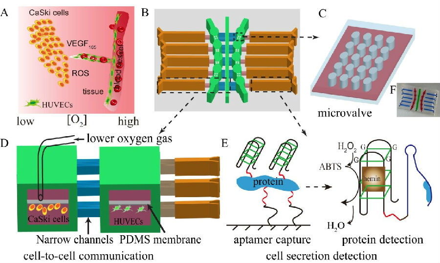
Recently, Prof. Jin-Ming Lin and his group from Department of Chemistry, Tsinghua University, have developed a multifunctional microfluidic platform with integration of oxygen-gradient microenvironment simulation, cell co-culture and cell metastatic VEGF165 on-line monitoring in one chip, which can enable to the study of endothelium-tumor interaction and tumor neovascular as well as cell migration. The results suggested that oxygen microenvironment imposes different impacts on the migration behavior and cell metabolism between endothelium and tumor cells: at 5% oxygen level, tumor cells presented stronger migration ability because of the better tolerance of low level oxygen, which may be corresponding to the in vivo tumor invasion under hypoxia condition; for 15% oxygen level, endothelium cell migrated faster than tumor, which would indicate the in vivo neovascular process. These results have provided significant experimental support for the study concerning cervical cancer occur and development. The work supported by National Natural Science Foundation (N.214350002) and the result of of oxygen-induced cell migration and on-line monitoring biomarkers modulation of cervical cancers on a microfluidic system has been published on Scientific Reports under Nature Publishing Group. (Sci. Rep., 2015, 5, 9643.)

The cell co-culture on a microfluidic chip with oxygen gradient in micro-scale
Prof. Jin-Ming Lin’s research group is dedicated to the development and application of chemiluminescence immunoassay kit for disease-related biomarker detection. Cooperation with a company has transformed some of the research results into great financial profit and social benefit, thus renowned Beijing Science and Technology Award in 2013. This research group also successfully developed cervical shedding cell preserve medium, and overcame the interference of blood and mucus from sample, greatly enhancing the slice production ratio with no pretreatment step. The perverse medium is able to extinguish pathogen in 15 min while preserving cells in intact morphology. With low cost, long effective duration and high positive detection rate, the technique was transferred to Minzhong Medical Technology Group Co., Ltd of Fujian Province in 2012 and now is widely applied among many hospitals around the country.